X-ray origins
Suddenly we could take photographs of our insides. And we loved it.
In 1899, a 14-year-old boy called Robert Luke went hunting with a friend on the Otago Peninsula. He used a ferret to chase a rabbit out of its hole and grabbed the animal’s back legs. His friend, exhibiting the lack of prefrontal cortex development 14-year-olds are known for, shot the rabbit, inadvertently sending pellets through
Robert Luke’s hand, too. When local doctor Henry de Lautour heard of the boy’s injuries, he thought of the technology he’d just imported—an early x-ray.
Robert presented his sore hand to the machine in front of an eager audience at the local church, and they enjoyed it so much that he was trotted out again for that evening’s Sunday School service.
For decades, an image of Robert’s hand was thought to be the oldest surviving x-ray in the country. But last year, a scan of a native frog in Canterbury Museum was pinned to 1896, pipping Robert by three years.
[Chapter Break]
Everyone alive today has grown up in a world where x-rays are taken for granted. But when the technology was first developed, the concept of seeing the inside of your body while you were still whole was a combination of titillating and terrifying. “It was shockingly novel, because skeletons had always been a symbol of death,” says Steven Muir, a medical physicist who has researched the history of x-rays in New Zealand.
The technology, invented in 1895 in Germany, travelled fast; by May 1896, it was in Dunedin. The potential seemed boundless. New Zealand newspapers published stories of hens being scanned for eggs, the prospect of full-body scans before marriage, and pointing rays at the ground to look for gold.
At first, the machines made for thrilling public displays. People scanned umbrellas, purses, the rings on their hands, plates of food. Robert Turnbull (brother of Alexander) imported an x-ray tube into Wellington and displayed it at his house on The Terrace. “The current is switched on, the glass globe lights up with a pale green glow, and the sitter wonders if he will emerge from the operation alive,” reads a report in the Evening Post.
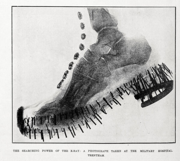
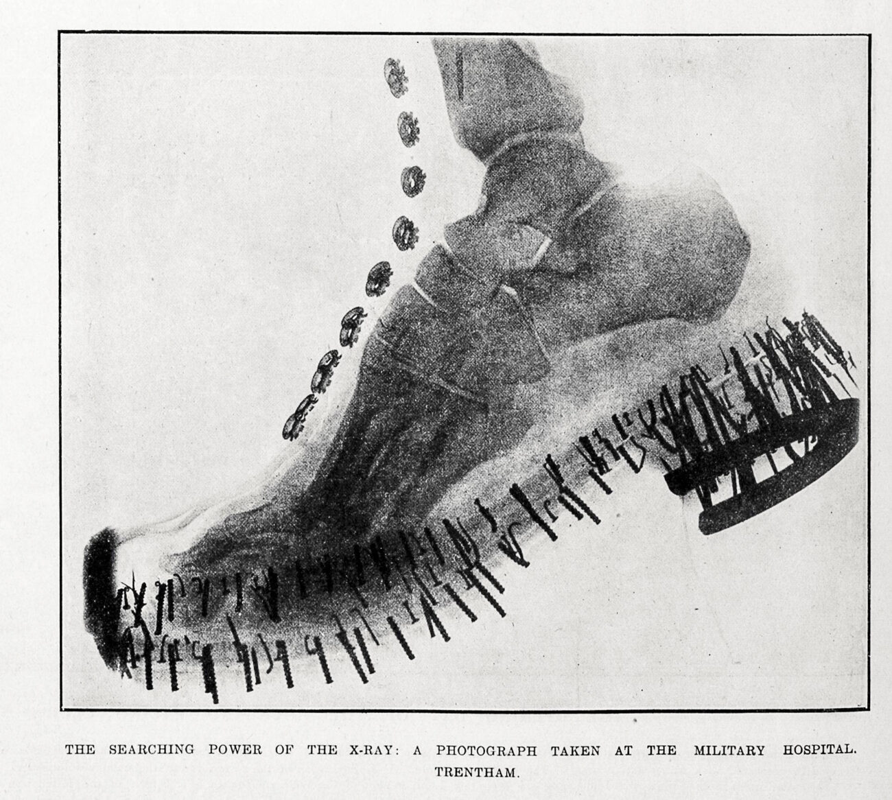
An exposure of one minute was enough to produce an image of a foot which, “greatly to the owner’s disgust, showed unmistakable evidences of his having worn tight boots”. The image hasn’t survived (the picture here is from the military hospital at Trentham) but you can imagine the revelation: boots were no longer impenetrable.
The harms of exposure were becoming clear, too. Unlike modern x-rays, where operators and tubes are shielded by lead, early devices sprayed rays out in all directions. In 1986, the Evening Post said that an English x-ray operator “lost most of his finger-nails and the skin of one hand”. A 1909 piece in New Zealand Graphic (no relation) said x-ray operators could become “living death personified”. “The tissues become corroded and life is only bearable under the influence of opiates.”
De Lautour tried to limit these dangers with some lead shielding, and he stood back from the machine at public displays, but many people were exposed to radiation in the early flush of x-ray enthusiasm, Muir says.
Robert Luke didn’t know it in 1899, but the display of his shot-riddled hand was the end of an era. Just four years after their invention, x-rays were becoming essential medical technology, but they typically no longer drew crowds; public attention was shifting to another exciting import, cinematography.
Revelations of bones are limited to radiology departments now, and if there is a thrill, it’s individual: There are my bones, and that is the machine that can see them.