Deep vision
A new type of underwater camera sees more than ever before.
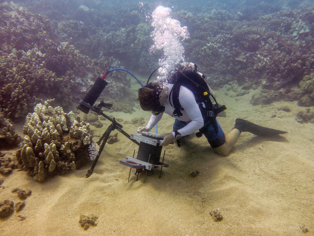
Dive photography stepped up a notch when a United States diver built a camera that can record animals on the seafloor as tiny as a tenth the size of a human hair.
The camera, designed by University of California San Diego PhD student Andrew Mullen, can capture detail in the field previously possible only in the lab—such as single-celled algae just 10 micrometres across that live in coral—revealing tiny interactions that together influence the marine ecosystem.
The camera uses a soft lens that focuses the way your eye does, by changing shape, and a tripod for placement on the seafloor. With 2.2 micrometres its maximum resolution, it uses ‘focal stacks’ to record animals that have a deeper 3D structure.
Leaving the camera in place overnight has revealed ‘coral polyp kissing’ for the first time in the natural environment, where polyps of the same colony periodically embraced each other throughout the night, perhaps as a way to swap materials. The team also placed corals from different areas together, and overnight footage captured them ejecting extensions of their gut to digest each other.